Introduction
Sternum, a part of rib cage, is located in center of the chest and protects many vital organs of the body like heart and lungs. It has three parts: Manubrium, body and Xiphoid process. During development of fetus sternum cartilage is formed by two bars which merge with each other towards the eighth week of gestation forming the manubrium and the body of the sternum.1
Any failure in this developmental process results in sternal variations and anomalies.2 A partial defect in the meeting of cartilage bars can cause holes to form in the sternum. Some segments are sometimes formed from more than one center, which varies in number and position. The first piece may therefore have two, three, or even six centers. When two are present, they are generally located one above the other, the upper being the larger; the second piece rarely has more than one center; the third, fourth and fifth pieces are often formed from two laterally placed centers, the irregular union of which explains the rare occurrence of the sternal foramen or the vertical fissure that occasionally intersects this part of the bone. Overall incidence of sternal foramen by Babinski et al. was 16.6%,3 Stark 4.3% in chest computed tomography,4 Cooper et al. 6.7%in autopsy cases.5 Mostly asymptomatic, incidentally found in X-ray which may be confused with gunshot wounds or lytic lesions.6 Sternum foramen may be misinterpreted as acquired lesions such as gunshot wound, fracture, lytic lesions, etc. Sternal foramen leaves the lung, heart and large vessels unprotected while performing invasive procedures such as bone marrow aspiration,7 acupuncture leading to life-threatening complications such as pneumothorax and cardiac tamponade.
Materials and Methods
The present study was carried using sternal bones removed during autopsy on a total of 350 cases above the age of 30 years at the Department of Forensic Medicine, Lady Hardinge Medical College, New Delhi. After removal of tissue and maceration, the sternum was examined for any kind of variation in its morphology. Steps of maceration:
Sterna were removed from the cadavers by sectioning the costal cartilages just besides the costo-chondral junction.
The sterna thus collected were marked, numbered, and then put in a water bath containing solution of sodium hypo-chloride for a week for maceration.
The sternum was then cleaned and examined intermittently to look for maceration on the 3rd, 5th and 7thdays.
After maceration and cleaning all the remains of the muscle and ligaments the sterna were dried at room temperature.
The morphometric parameters were expressed in mm. All macroscopic measurements were made to the nearest 0.01mm using digital calipers, and no distinction was made as to ethnical and sexual dimorphism of the dry bones. For statistical analysis of the frequency and diameter of sternal foramen, the software SPSS version 16 was used.
Results
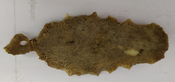
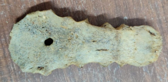
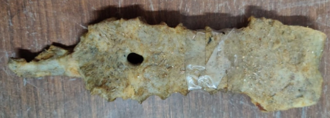
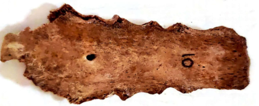
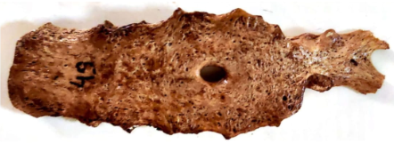
Sternal foramina were observed as single midline defects in inferior half of the body of sternum intwenty-three out of total three hundred fifty sternum studied,incidence being 6.57%. Size of sternal foramina width ranged between 6.50 and 7.72 mm with a mean of 7.70 mm. Sternal foramen mean distance from sternal notch was 107.41 mm, and distance from the lower end of the sternum was 36.57 mm. The vertical and transverse diameters were measured. In each foramen the vertical diameter was noted to be more than the transverse diameter.
Discussion
The current study has established an incidence of 6.57% for sternal foramina. This is similar to the findings of Cooper et al.,5 Moore et al.,8 Alkan and Savas.9 Cooper et al. have reported an incidence of 6.7% in an autopsy study. Moore et al. have found an incidence of 6.6% whereas the incidence reported by Alkan and Savas is 5.1%. Findings of the study are comparable to the 4.3% and 4.5% incidence observed by Stark4 and Yekeler et al.10 respectively. Peuker et al.11 have also reported an incidence between 5% and 8% in autopsy studies. Among Indian studies, Shivkumar et al.12 have reported a comparable incidence of 7%. Another study by Chaudhari et al.2 has revealed an incidence of 4.1%. However, higher incidence of 13.8% and 16.6% has been reported by El-Busaid et al.13 and Babinski et al.3 respectively. Morphometry of sternal foramen in the current study has established similarity with the finding of Yekeler et al.10 They have reported the size of sternal foramina ranging from 2 mm and 16 mm. Findings of the current study fall in the same range with the maximum diameter being the vertical one.
The ossification centers of manubrium and sternal body form on a cartilaginous plate on either side of the midline in cranio-caudal direction, starting from the 5th month of prenatal life to shortly before birth. The ossification centers usually fuse in manubrium before birth. The sternum begins to develop on either side of the midline through mesenchymal bands. These later fused to form a wide triangular manubrium. The slender xiphoid process is continuous with the lower end of the body at the xiphisternal joint.
Union between the sternebral centers begins at puberty and continues upward from below. Thus, the arrangement and number of ossification sternal centers vary depending on the level of completeness and time of sternal plate fusion. The manubrium is ossified from one to three centers which appear in the 5th fetal month. First and second sternebrae ossify from the single center around 5th fetal month. For third and fourth sternebrae, a pair of ossification centers for each appears in 5th and 6th fetal month respectively. The xiphoid process begins to ossify after birth around 3rd year. Sternal foramen is the result of incomplete fusion.14 For medical practitioners, a sound awareness of sternal variations and irregularities is beneficial in bone marrow screening, radiology documentation, acupuncture practice for improved diagnosis and care so as not to confuse them with pathological conditions. The morphological variants of the xyphoid process are important for cardiothoracic surgeons, doctors and radiologists to avoid misdiagnosis. When marking pathological disorders such as traumatic fissures, fractures and osteolytic lesions in sternum cross-sectional imagery, imaging appearances of sternal and xiphoid variance and abnormalities should be kept in mind. Sternal or xiphoid foramina may pose a great hazard during sternal puncture, due to inadvertent injury to heart or great vessels. Variations in length or size of xiphoid process are likely to pose problems for clinicians such as very rare long and enlarged xiphoid process may decrease the xiphisternal angle and more likely to develop xiphoidalgia or may be mistaken for an epigastric mass.15 In addition, an unusually longer xiphoid process with sharp bifid ends may misguide practitioners for a bony landmark during cardiopulmonary resuscitation to determine where chest compressions are to be administered or may be mistaken for fractures during imaging.
The incidence of the sternal foramen is 4.3% in living and 6.7% in autopsy cases.16 Most of the foramina were located in the lower part of the body. Mostly, sternal foramen is an isolated developmental anomaly but sometimes may be associated with accessory fissures on left lung. Presence of xiphoid foramen can lead to misinterpretation and wrong conclusions as bullet injuries which have serious consequences in addition the knowledge of this occurrence is important to avoid serious heart injury by needle insertion especially since this area holds a commonly used acupuncture point and sternal puncture. However, in case such confusion prevails in medicolegal autopsy and sternal foramen are misjudged as gun-shot wounds or antemortem trauma, the definitive diagnosis of sternal foramen can be made on close and meticulous examination. Careful examination will reveal that an alleged sternal foramen will mostly be a midline defect in the lower half of the mesosternum with a smooth margin covered with cortical bone as opposed to a gun-shot wound showing bevelled outline with fracture lines surrounding the wound and the absence of cortical bone on the edges. Similarly, the presence of typical erosion by insects and animal claw or teeth marks should be countered by an antemortem trauma assumption. In the event of such confusion, careful pathological and biochemical investigations should be carried out to exclude osteolytic conditions. From a medicolegal perspective, identifying the morphological anomalies or variations of xiphoid process aid in the individualization process by serving as points of similarity when their occurrence has been recorded antemortem. In addition, their antemortem records in the form of previous X-rays make important data for skeletonized remains to be identified.
Conclusion
The sternal foramen and bifid xiphoid process are the most common variants and are mostly asymptomatic. The sternal foramen can be mistaken as acquired lesion, like a gunshot wound. Sternum is closely related to mediastinal structures, the presence of sternal foramen makes the lung, heart and large vessels vulnerable while performing invasive procedures; therefore, it is prudent to take X-rays to rule out such sternum failure; before performing intrusive procedures such as bone marrow aspiration, acupuncture leading to life-threatening impediments such as pneumothorax. Sternal foramen is a variation in anatomy due to defective sternum ossification. Knowledge of this anomaly is very important for medical staff, radiologists and acupuncture, as accidental pericardium penetration by the needle through such a defect can lead to pericardial effusion or cardiac tamponade during bone marrow aspiration and acupuncture. It is recommended to use proper Multi Detector C T imaging to avoid such hazardous and life-threatening complications. Forensic pathologists should be meticulous enough to rule out errors in determining the nature and cause of death in worrying cases as sternal foramina is highly likely to be confused with gun-shot wounds or traumatic antemortem injury.


